
撰文|郭洋(2024级博士研究生)
审核|孙少聪
Speaker Bio

Manolis Pasparakis is Professor at the Institute for Genetics, University of Cologne, where he leads a program focused on cell death and inflammation. His laboratory has been one of the major forces in redefining regulated cell death from a largely cell-autonomous endpoint into a driver of tissue pathology, barrier failure, and chronic inflammation.
Abstract
The central tenet of this lecture is that caspase-8 functions as a homeostatic checkpoint, determining whether inflammatory receptor signaling remains compatible with tissue integrity or is diverted toward apoptosis, necroptosis, pyroptosis, or integrated lytic outcomes. Consequently, the field has moved away from a narrow "cell death mechanism" framework toward one centered on tissue homeostasis. In barrier tissues, the decisive question is not whether cells die, but rather how they die, under which checkpoint constraints, and with what consequences for the surrounding tissue.
Work over the past decade has shown that caspase-8 restrains the cytotoxic activity of RIPK1 via proteolytic cleavage and coordinated checkpoint regulation, terminates cytotoxic signalling complexes through cleavage events, interacts functionally with cFLIP, modulates inflammasome activation and downstream inflammatory outputs, and influences cytokine production independently of its canonical apoptotic role. These functions are especially important in the intestine, skin and liver, where aberrant death programmes convert epithelial stress into chronic inflammation. Recent studies further show that endogenous nucleic-acid sensing, ZBP1-driven signalling, STING activation, the co-occurrence of apoptosis, necroptosis and pyroptosis within a cell population, and non-apoptotic caspase-8 outputs broaden the mechanistic landscape far beyond the classical apoptosis–necroptosis dichotomy. Collectively, the evidence positions caspase-8 as a central molecular switch linking cell fate, inflammatory amplification and long-term organ integrity.
Keywords
Caspase-8; RIPK1; necroptosis; pyroptosis; tissue homeostasis; inflammation; ZBP1; barrier tissues.
Caspase-8 as a cell-fate checkpoint rather than a simple apoptotic initiator
Caspase-8 serves as the core regulator of the TNFR1 signaling decision node. After TNF ligation, Complex I forms at the receptor and contains RIPK1, TRADD, TRAF2, cIAPs, LUBAC and associated kinase modules that support NF-κB and MAPK signalling. Under homeostatic conditions, ubiquitylation and phosphorylation of RIPK1, together with NEMO/IKK-, TAK1- and TBK1/IKKε-dependent checkpointing, keep RIPK1 in a pro-survival or at least non-cytotoxic state. When these constraints fail, RIPK1 forms cytosolic death-inducing complexes with FADD and caspase-8, leading to apoptosis, necroptosis, or mixed inflammatory outcomes (Fig1).
Within this framework, caspase-8 catalytic activity and its cleavage of RIPK1 are not redundant details; they function as critical physiological brakes on inflammatory pathology. Catalytically competent caspase-8, especially in a complex with cFLIPL, suppresses RIPK3-dependent necroptosis during development. Thus, the most accurate current description is that caspase-8 sits at a fate checkpoint: enough activity can dismantle death-inducing complexes and cleave key substrates such as RIPK1, whereas loss of that activity destabilizes the checkpoint and permits cytotoxic signalling to escalate into apoptosis, necroptosis or inflammasome-linked death programmes. Caspase-8-mediated cleavage of RIPK1 is essential for limiting aberrant apoptosis and necroptosis, confirming that this cleavage acts as a non-redundant in vivo brake rather than a minor biochemical detail. Catalytically inactive caspase-8 can redirect signalling toward ASC-, caspase-1- and pyroptosis-associated pathology. These studies effectively established caspase-8 as a molecular switch among apoptosis, necroptosis and pyroptosis.
Recently, N4BP1 has been identified as a physiologically relevant substrate of caspase-8 whose cleavage integrates TLR, TRIF and TNF signalling. Therefore, Caspase-8 is not merely 'pro-death' or 'anti-necroptotic'; it also participates directly in licensing inflammatory cytokine production. In other words, caspase-8 can shape immune responsiveness without necessarily committing the cell to terminal death, a point that helps explain why human CASP8 deficiency combines immunodeficiency with dysregulated inflammation rather than resembling a pure apoptosis defect.
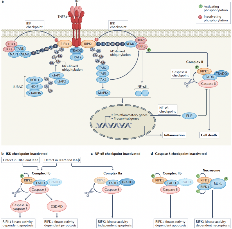
Fig1. Caspase-8 as a central checkpoint coordinating cell fate decisions downstream of TNFR1 signaling
From regulated cell death to tissue homeostasis in barrier organs
The most informative biological question is not simply how a cell dies, but how death signaling is prevented, redirected, or tolerated to preserve tissue homeostasis under inflammatory stress. In the intestine, skin and liver, the relevant biological unit is not the individual dying cell but the barrier or parenchymal system. Caspase-8 and RIPK1 are therefore best understood as key regulators of tissue architecture, because they determine whether ordinary inflammatory cues are absorbed and resolved, or instead translated into barrier rupture, microbial translocation, stromal activation and self-amplifying inflammation.
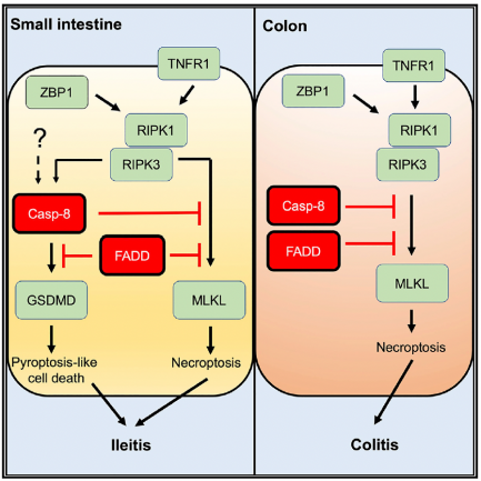
Genetic evidence in the intestinal epithelium underscores an integrated cell death network regulating tissue homeostasis. Epithelial FADD and caspase-8 prevent chronic intestinal inflammation and terminal ileitis by suppressing RIPK3- and TNF-driven necroptosis. Upstream, RIPK1 maintains epithelial integrity by restraining both apoptosis and necroptosis, while NEMO acts through both NF-κB-dependent and -independent pathways to suppress RIPK1-driven cell death. Crucially, FADD and caspase-8 orchestrate gut homeostasis by concurrently constraining MLKL-mediated necroptosis and GSDMD-mediated pyroptosis, defining intestinal pathology as an integrated death-network dysfunction rather than a lineage-restricted necroptosis phenotype (Fig2).
In skin, the inflammatory outcome of cell death is dictated by context and regulated modalities. Epidermal deficiency in FADD or caspase-8 triggers severe inflammatory dermatitis driven by RIPK3/MLKL-dependent necroptosis. Similarly, loss of RIPK1 scaffold function induces pathology by unleashing ZBP1-driven necroptosis, a process activated through the sensing of endogenous nucleic acids. Consequently, inflammatory dermatitis in these models represents a checkpoint failure that converts intrinsic epithelial stress into immunogenic cell death, rather than a generic response to tissue damage, directly coupling cell death control to innate immune surveillance.
In the liver, loss of NEMO in hepatocytes drives RIPK1 kinase-dependent apoptosis, which leads to steatohepatitis, compensatory proliferation, and ultimately hepatocellular carcinoma. This pathology demonstrates that apoptosis can act as a chronic inflammatory and tumor-promoting driver when cell loss is functionally coupled to tissue regeneration and innate immune activation. Therefore, organ-level pathogenesis depends not only on lytic cell death (such as necroptosis or pyroptosis) but also on non-lytic apoptosis, provided its rate, persistence, or clearance dynamics are altered.
Collectively, these studies indicate that tissue pathology arises not simply from excessive cell death, but from the inappropriate execution of cell death programs within anatomically specialized tissues.
 |
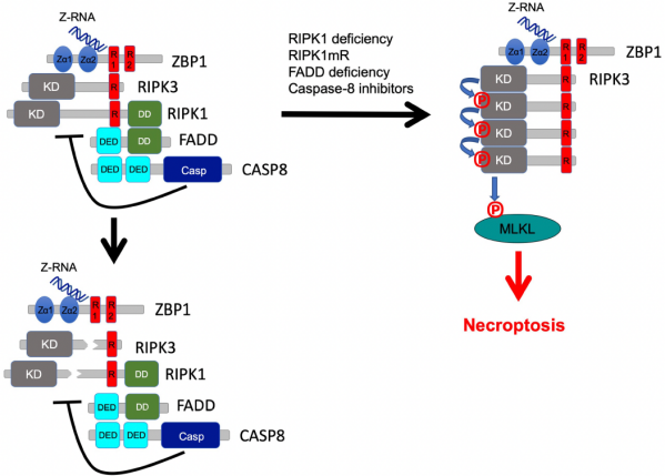 |
Fig2. Tissue-specific functions of caspase-8 and RIPK1 in maintaining epithelial homeostasis
Cell death as a trigger of inflammation
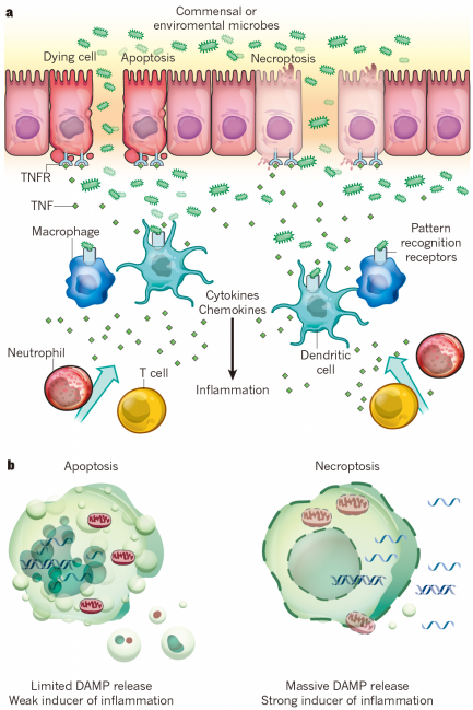
Cell death actively initiates and amplifies tissue inflammation rather than serving as a passive consequence of injury. While lytic modalities (necroptosis and pyroptosis) directly release DAMPs and intracellular antigens, death-induced inflammation in vivo fundamentally stems from disrupted barrier permeability, altered stromal signaling, shifted cytokine gradients, and enhanced immune recruitment (Fig3).
Necroptosis exemplifies this inflammatory capacity, particularly through the activation of endogenous nucleic-acid sensing. When RIPK1 or caspase-8 checkpoints fail, ZBP1-mediated sensing of endogenous Z-form nucleic acids triggers necroptosis and localized tissue pathology; this mechanism is further supported by a cGAS-STING-ZBP1 signaling axis that drives TNFR1-independent necroptosis.
Notably, apoptosis is immunologically silent only under homeostatic conditions, where rapid efferocytosis clears dying cells.; however, it becomes pro-inflammatory and oncogenic when the death rate overwhelms clearance capacity, when secondary necrosis ensues, when barrier integrity is compromised, or when apoptotic signaling couples to cytokine processing (as demonstrated in SHARPIN-deficient skin disease and NEMO-deficient hepatocarcinogenesis).
Furthermore, the death machinery can initiate inflammatory programs prior to complete execution. Upstream ripoptosome-associated caspase-8 can directly process IL-33 to drive type 2 immunity, while TAK1 inhibition promotes caspase-8-dependent GSDMD cleavage.
Consequently, tissue pathogenesis is ultimately dictated not by cell death but by abnormal modes of death, anatomical location, and kinetics, in which checkpoint failure transforms homeostatic cell turnover into a sustained inflammatory cascade.
 |
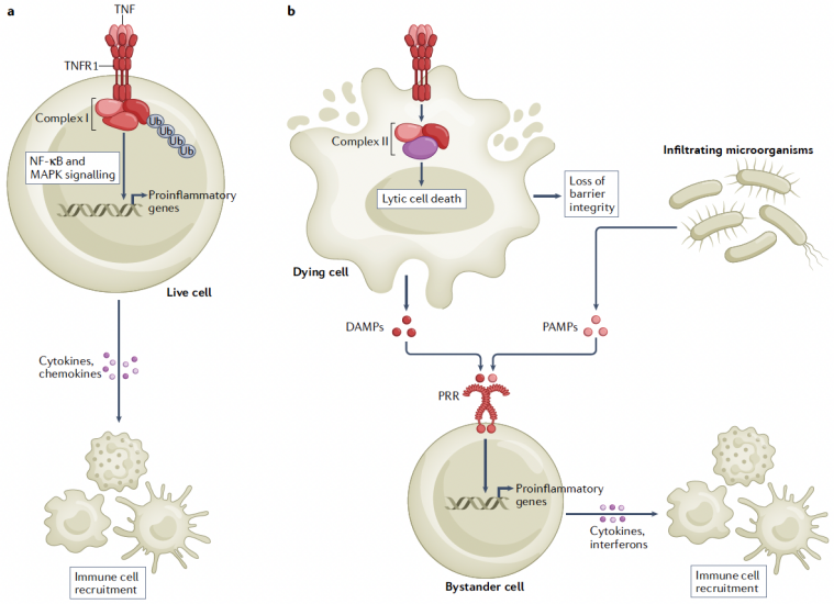 |
Fig3. How dying cells trigger inflammation in vivo
Integrated death networks beyond the classical apoptosis–necroptosis dichotomy
Over the past decade, regulated cell death has been redefined as an integrated network rather than a set of insulated pathways. Caspase-8 sits at the center of this paradigm shift. When canonical apoptosis and MLKL-mediated necroptosis are constrained, caspase-8 signaling shifts toward ASC-dependent inflammasome assembly, caspase-1 activation, and pyroptosis.
The distinction between apoptosis and pyroptosis is functionally blurred, as caspase-8 can engage either pathway depending on the signaling context. Under conditions like TAK1 blockade, caspase-8 directly cleaves GSDMD to drive inflammatory cell death, converting an apoptotic driver into a pyroptotic executioner depending on pathogen and cytokine context.
Cell death machinery can act heterogeneously within a cell population: apoptosis, necroptosis and pyroptosis often coexist rather than being driven by one distinct pathway. Innate sensors such as ZBP1, AIM2 and Pyrin can form multiprotein complexes that recruit ASC, caspase-1, caspase-8, RIPK1, RIPK3 and FADD. In these platforms, caspase-8 acts as a structural and enzymatic bridge linking death receptors, inflammasomes and RIP kinases.
The ZBP1 axis further broadens this network. Activated by endogenous Z-form nucleic acids and boosted by STING signaling, ZBP1 converts nucleic-acid sensing into necroptotic inflammation. Importantly, caspase-8 deficiency does not merely unleash necroptosis; it reconfigures interferon-driven transcriptional states to enhance necroptotic sensitivity.
Finally, caspase-8 operates beyond cell death execution. In severe SARS-CoV-2 infection and metabolic dysfunction-associated steatohepatitis (MASH), caspase-8 actively drives tissue inflammation and fibrosis through non-apoptotic signaling pathways. Thus, the physiological question is no longer 'which isolated pathway is active,' but rather 'which checkpoint has failed and which signaling outlet has opened' (Fig4).
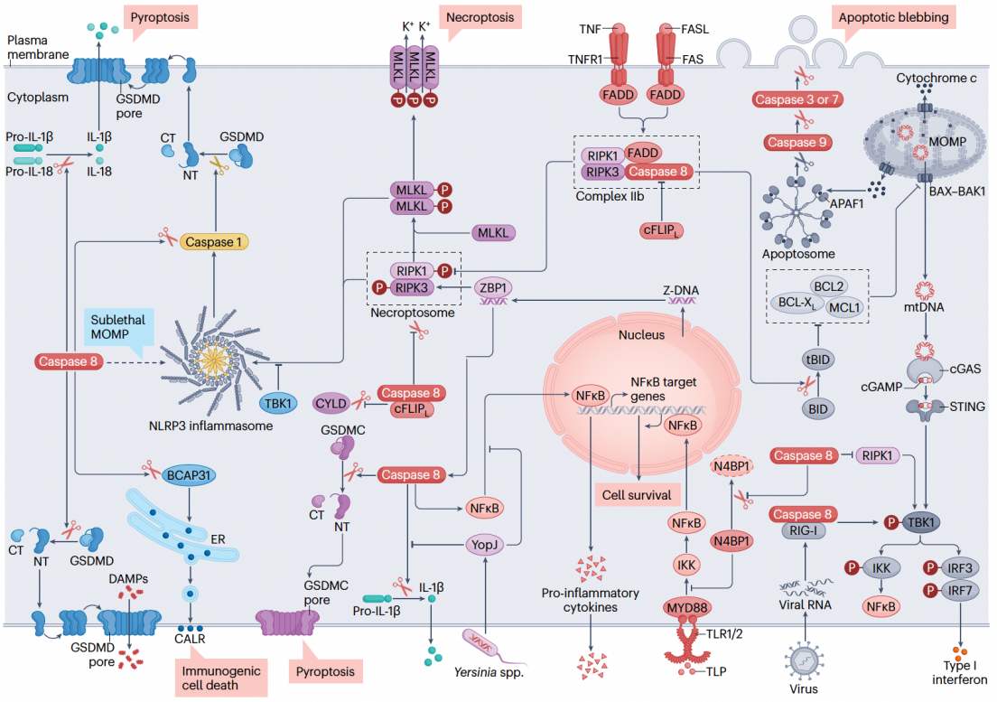
Fig4. Integrated death networks centered on caspase-8
References
1. Kondylis V, Kumari S, Vlantis K, Pasparakis M. The interplay of IKK, NF-κB and RIPK1 signaling in the regulation of cell death, tissue homeostasis and inflammation. Immunol Rev. 2017;277(1):113–127. doi:10.1111/imr.12550.
2. Pasparakis M, Vandenabeele P. Necroptosis and its role in inflammation. Nature. 2015;517(7534):311–320. doi:10.1038/nature14191.
3. Welz PS, Wullaert A, Vlantis K, et al. FADD prevents RIP3-mediated epithelial cell necrosis and chronic intestinal inflammation. Nature. 2011;477(7364):330–334. doi:10.1038/nature10273.
4. Günther C, Martini E, Wittkopf N, et al. Caspase-8 regulates TNF-α-induced epithelial necroptosis and terminal ileitis. Nature. 2011;477(7364):335–339. doi:10.1038/nature10400.
5. Dannappel M, Vlantis K, Kumari S, et al. RIPK1 maintains epithelial homeostasis by inhibiting apoptosis and necroptosis. Nature. 2014;513(7516):90–94. doi:10.1038/nature13608.
6. Takahashi N, Vereecke L, Bertrand MJM, et al. RIPK1 ensures intestinal homeostasis by protecting the epithelium against apoptosis. Nature. 2014;513(7516):95–99. doi:10.1038/nature13706.
7. Vlantis K, Wullaert A, Polykratis A, et al. NEMO prevents RIP kinase 1-mediated epithelial cell death and chronic intestinal inflammation by NF-κB-dependent and -independent functions. Immunity. 2016;44(3):553–567. doi:10.1016/j.immuni.2016.02.020.
8. Kondylis V, Polykratis A, Ehlken H, et al. NEMO prevents steatohepatitis and hepatocellular carcinoma by inhibiting RIPK1 kinase activity-mediated hepatocyte apoptosis. Cancer Cell. 2015;28(5):582–598. doi:10.1016/j.ccell.2015.10.001.
9. Lin J, Kumari S, Kim C, et al. RIPK1 counteracts ZBP1-mediated necroptosis to inhibit inflammation. Nature. 2016;540(7631):124–128. doi:10.1038/nature20558.
10. Oberst A, Dillon CP, Weinlich R, et al. Catalytic activity of the caspase-8–FLIP<sub>L</sub> complex inhibits RIPK3-dependent necrosis. Nature. 2011;471(7338):363–367. doi:10.1038/nature09852.
11. Newton K, Wickliffe KE, Dugger DL, et al. Cleavage of RIPK1 by caspase-8 is crucial for limiting apoptosis and necroptosis. Nature. 2019;574(7778):428–431. doi:10.1038/s41586-019-1548-x.
12. Lalaoui N, Boyden SE, Oda H, et al. Mutations that prevent caspase cleavage of RIPK1 cause autoinflammatory disease. Nature. 2020;577(7788):103–108. doi:10.1038/s41586-019-1828-5.
13. Newton K, Wickliffe KE, Maltzman A, et al. Activity of caspase-8 determines plasticity between cell death pathways. Nature. 2019;575(7784):679–682. doi:10.1038/s41586-019-1752-8.
14. Fritsch M, Günther SD, Schwarzer R, et al. Caspase-8 is the molecular switch for apoptosis, necroptosis and pyroptosis. Nature. 2019;575(7784):683–687. doi:10.1038/s41586-019-1770-6.
15. Gitlin AD, Heger K, Schubert AF, et al. Integration of innate immune signalling by caspase-8 cleavage of N4BP1. Nature. 2020;587(7833):275–280. doi:10.1038/s41586-020-2796-5.
16. Orning P, Weng D, Starheim K, et al. Pathogen blockade of TAK1 triggers caspase-8-dependent cleavage of gasdermin D and cell death. Science. 2018;362(6418):1064–1069. doi:10.1126/science.aau2818.
17. Demarco B, Grayczyk JP, Bjanes E, et al. Caspase-8-dependent gasdermin D cleavage promotes antimicrobial defense but confers susceptibility to TNF-induced lethality. Sci Adv. 2020;6(47):eabc3465. doi:10.1126/sciadv.abc3465.
18. Schwarzer R, Jiao H, Wachsmuth L, Tresch A, Pasparakis M. FADD and caspase-8 regulate gut homeostasis and inflammation by controlling MLKL- and GSDMD-mediated death of intestinal epithelial cells. Immunity. 2020;52(6):978–993.e6. doi:10.1016/j.immuni.2020.04.002.
19. Jiao H, Wachsmuth L, Kumari S, et al. Z-nucleic-acid sensing triggers ZBP1-dependent necroptosis and inflammation. Nature. 2020;580(7803):391–395. doi:10.1038/s41586-020-2129-8.
20. Devos M, Tanghe G, Gilbert B, et al. Sensing of endogenous nucleic acids by ZBP1 induces keratinocyte necroptosis and skin inflammation. J Exp Med. 2020;217(7):e20191913. doi:10.1084/jem.20191913.
21. Christgen S, Zheng M, Kesavardhana S, et al. AIM2 forms a complex with pyrin and ZBP1 to drive PANoptosis and host defence. Nature. 2021;600(7889):415–419. doi:10.1038/s41586-021-03875-8.
22. Lafont E, Draber P, Rieser E, et al. TBK1 and IKKε prevent TNF-induced cell death by RIPK1 phosphorylation. Nat Cell Biol. 2018;20(12):1389–1399. doi:10.1038/s41556-018-0229-6.
23. van Loo G, Bertrand MJM. Death by TNF: a road to inflammation. Nat Rev Immunol. 2023;23(5):289–303. doi:10.1038/s41577-022-00792-3.
24. Yuan J, Ofengeim D. A guide to cell death pathways. Nat Rev Mol Cell Biol. 2024;25(5):379–395. doi:10.1038/s41580-023-00689-6.
25. Beltrán-Visiedo M, Soler-Agesta R, Sarosiek KA, Green DR, Galluzzi L. Regulation of inflammatory processes by caspases. Nat Rev Mol Cell Biol. 2025;26:884–901. doi:10.1038/s41580-025-00869-6.
26. Lehle AS, Farin HF, Marquardt B, et al. Intestinal inflammation and dysregulated immunity in patients with inherited caspase-8 deficiency. Gastroenterology. 2019;156(1):275–278. doi:10.1053/j.gastro.2018.09.041.
27. Wang X, Moore MP, Shi H, et al. A non-apoptotic caspase-8–meteorin pathway in hepatocytes promotes MASH fibrosis. Nat Metab. 2025;7:2067–2082. doi:10.1038/s42255-025-01355-1.
28. Kelepouras K, et al. STING induces ZBP1-mediated necroptosis independently of TNFR1 and FADD. Nature. 2025. doi:10.1038/s41586-025-09536-4.


 北京市丰台区右安门外西头条10号
 010-86738999
 cimr@cimrbj.ac.cn


 En
En

